- Dawes GS, Milne ED, Mott JC, Widdicombe JG (1953) The patency of the
ductus arteriosus after birth. J Physiol. 1953 Oct;122(1):37-8P. - Dawes GS, Milne ED, Mott JC, Widdicombe JG.The closure of the foramen
ovale after birth. J Physiol. 1953 Oct;122(1):38P. - Born GV, Dawes GS, Mott JC, Widdicombe JG (1954) Changes in the heart
and lungs at birth. Cold Spring Harbor Symposia on Quantitative Biology
1954;19:102-8. - Gunther M (1957) The transfer of blood between baby and placenta in the
minutes after birth. Lancet. 1957 Jun 22;272(6982):1277-80. - Haselhorst G (1929) Uber Art und Dauer der Blutstromung in den
Nabelschnurgefassen post partum. Z Geburtshilfe Gynakol 96:487-499
(Zeitschrift für Geburtshilfe und Gynäkologie) - Allmeling A (1930) Die Gewichtszunahme von Neugeborenen infolge
postnataler Transfusion. Zentralbl Gynakol 54:850-860. (Zentralblatt für
Gynäkologie) - Jaykka S (1957) Capillary erection and lung expansion; an experimental
study of the effect of liquid pressure applied to the capillary network of
excised fetal lungs. Acta Paediatr. 1957 Jan;46(suppl 112):1-91. - Orth J (1875) Ueber das Vorkommen von Bilirubinkrystallen bei
neugebornen Kindern. Archiv für pathologische Anatomie und Physiologie
und für klinische Medicin 63:447-462 - Schmörl G (1904) Zur Kenntnis des Ikterus neonatorum, insbesondere der
dabie auftretenden Gehirn veränderungen. Verhandlung der deutschen
pathologischen Gesellschaft 6:109-115. - Zimmerman HM and Yannet H (1933). Kernicterus: jaundice of the nuclear
masses of the brain. American Journal of Diseases of Children, 45, 740-
759. - Lucey JF, Hibbard E, Behrman RE, Esquival FO, Windle WF (1964)
Kernicterus in asphyxiated newborn monkeys. Experimental Neurology 9:
43-58.
Postnatal transfer of blood from placenta to the
newborn infant, Gunther (1957)
Apgar's newborn scoring system seems clearly
associated with the vogue of early clamping of the
umbilical cord -- done with the aim of transferring the
infant to a specialist and maintaining a sterile field
for prompt suturing of incisions made for episiotomy
or cesarean delivery. Meanwhile, advocates of what
Apgar referred to as "slow birth" continued a long
tradition of measuring the amount of placental blood
an infant got if the cord was left unclamped until
pulsations in it ceased.
Pulsations continue as long as the valves in the
infant's heart direct blood to the placenta through
the umbilical arteries, and cease when pulmonary
respiration is fully established with closure of the
foramen ovale and ductus arteriosus in the heart
(Dawes et al. 1953).
Changes in circulation through the heart and lungs
were determined in research with newborn lambs.
These experiments did involve "tying the cord,"
which might explain the finding of a pattern of
"neonatal circulation" intermediate between that of
the fetus and that of the adult. Born et al. (1954)
found the ductus arteriosus begins to close within 5
to 15 minutes of pulmonary ventilation with
continuing constriction for several minutes, but
remained partially patent for 12 hours or more. The
foramen ovale, on the other hand, closes within a
minute following birth, forcing circulation to the lungs.
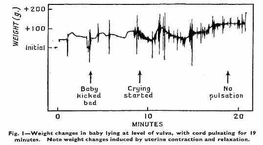
Gunther (1957) weighed infants for up to 20 minutes
after birth, with the umbilical cord intact allowing
ongoing placental circulation. Fluctuations in weight
occurred in response to uterine contractions,
elevation of the baby above or below the mother's
uterus, and pulsations of the cord.
Most interesting of Gunther's findings were the
annotations of the weight gain/loss tracings. In
figure 1 postnatal activity and weight profile are
shown for a baby who started crying only 9 minutes
after birth, and with pulsations of the cord continuing
for 19 minutes after birth. What would have been
the Apgar scores and fate of this child had the cord
been cut within the first minute after birth?
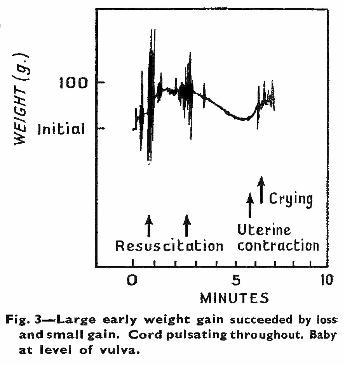
Likewise, in figure 3, resuscitation was started within
one minute on an infant described as "slow to cry."
Crying began more than 6 minutes after birth
following a uterine contraction and additional weight
gain from placental blood. Pulsations of the cord
continued throughout the 10 minute interval shown
in the graph. This would surely have been another
infant described as severely depressed by Apgar,
and with an ominous outcome.
Gunther commented, "These records confirm once
more that, if the cord is left untied, a baby will usually
increase his blood volume by a significant amount."
She compared her findings to those of Haselhorst
(1929) and Allmeling (1930), noting that placental
transfusion increased a newborn's weight by 0.8 to
4.7 percent, which (assuming blood volume is about
10 percent of an infant's weight) amounts to as much
or more than 40 percent of the baby's blood volume.
At this point in time, "pulmonary syndrome," later
referred to as hyaline membrane syndrome, was a
major concern, and quite widely attributed to the new
vogue of early umbilical cord clamping. Gunther
commented that even while pulsations of the cord
continued, cessation of placental transfusion was
often apparent, "as if a main reservoir had been
filled," and she cited the research of Jaykka (1957)
who determined that inflation of the lungs occurred
with increasing blood flow into the alveolar capillaries
- the shift of blood volume from the placenta to the
lungs.
newborn infant, Gunther (1957)
Apgar's newborn scoring system seems clearly
associated with the vogue of early clamping of the
umbilical cord -- done with the aim of transferring the
infant to a specialist and maintaining a sterile field
for prompt suturing of incisions made for episiotomy
or cesarean delivery. Meanwhile, advocates of what
Apgar referred to as "slow birth" continued a long
tradition of measuring the amount of placental blood
an infant got if the cord was left unclamped until
pulsations in it ceased.
Pulsations continue as long as the valves in the
infant's heart direct blood to the placenta through
the umbilical arteries, and cease when pulmonary
respiration is fully established with closure of the
foramen ovale and ductus arteriosus in the heart
(Dawes et al. 1953).
Changes in circulation through the heart and lungs
were determined in research with newborn lambs.
These experiments did involve "tying the cord,"
which might explain the finding of a pattern of
"neonatal circulation" intermediate between that of
the fetus and that of the adult. Born et al. (1954)
found the ductus arteriosus begins to close within 5
to 15 minutes of pulmonary ventilation with
continuing constriction for several minutes, but
remained partially patent for 12 hours or more. The
foramen ovale, on the other hand, closes within a
minute following birth, forcing circulation to the lungs.
Gunther (1957) weighed infants for up to 20 minutes
after birth, with the umbilical cord intact allowing
ongoing placental circulation. Fluctuations in weight
occurred in response to uterine contractions,
elevation of the baby above or below the mother's
uterus, and pulsations of the cord.
Most interesting of Gunther's findings were the
annotations of the weight gain/loss tracings. In
figure 1 postnatal activity and weight profile are
shown for a baby who started crying only 9 minutes
after birth, and with pulsations of the cord continuing
for 19 minutes after birth. What would have been
the Apgar scores and fate of this child had the cord
been cut within the first minute after birth?
Likewise, in figure 3, resuscitation was started within
one minute on an infant described as "slow to cry."
Crying began more than 6 minutes after birth
following a uterine contraction and additional weight
gain from placental blood. Pulsations of the cord
continued throughout the 10 minute interval shown
in the graph. This would surely have been another
infant described as severely depressed by Apgar,
and with an ominous outcome.
Gunther commented, "These records confirm once
more that, if the cord is left untied, a baby will usually
increase his blood volume by a significant amount."
She compared her findings to those of Haselhorst
(1929) and Allmeling (1930), noting that placental
transfusion increased a newborn's weight by 0.8 to
4.7 percent, which (assuming blood volume is about
10 percent of an infant's weight) amounts to as much
or more than 40 percent of the baby's blood volume.
At this point in time, "pulmonary syndrome," later
referred to as hyaline membrane syndrome, was a
major concern, and quite widely attributed to the new
vogue of early umbilical cord clamping. Gunther
commented that even while pulsations of the cord
continued, cessation of placental transfusion was
often apparent, "as if a main reservoir had been
filled," and she cited the research of Jaykka (1957)
who determined that inflation of the lungs occurred
with increasing blood flow into the alveolar capillaries
- the shift of blood volume from the placenta to the
lungs.
- Dawes GS et al. (1953) The
patency of the ductus
arteriosus after birth. - Dawes GS et al. (1953) The
closure of the foramen ovale
after birth. - Born GV (1954) Changes in
the heart and lungs at birth. - Gunther M (1957) The
transfer of blood between
baby and placenta in the
minutes after birth. - Haselhorst G (1929) Uber Art
und Dauer der Blutstromung
in den Nabelschnurgefassen
post partum. - Allmeling A (1930) Die
Gewichtszunahme von
Neugeborenen infolge
postnataler Transfusion. - Jaykka S (1957) Capillary
erection and lung expansion;
an experimental study of the
effect of liquid pressure
applied to the capillary
network of excised fetal lungs. - Orth J (1875) Ueber das
Vorkommen von
Bilirubinkrystallen bei
neugebornen Kindern. - Schmörl G (1904) Zur
Kenntnis des Ikterus
neonatorum, insbesondere
der dabie auftretenden
Gehirn veränderungen. - Zimmerman HM, Yannet H
(1933). Kernicterus: jaundice
of the nuclear masses of the
brain. - Lucey JF et al. (1964)
Kernicterus in asphyxiated
newborn monkeys.
Figures from Gunther (1957)