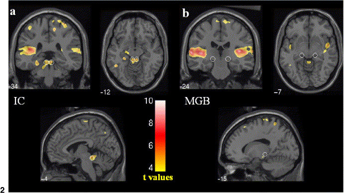
Figure 3: Blood flow in the human brain, revealed in fMRI scans. Functional MRI (fMRI) slices centered on
(a) the inferior colliculi (IC) left and (b) the medial geniculate bodies (MGB) right, indicated by white circles.
Note the prominent activity in the inferior colliculi.
From Budd TW, Hall DA, Goncalves MS, Akeroyd MA, Foster JR, Palmer AR, Head K, Summerfield AQ. Binaural specialisation in
human auditory cortex: an fMRI investigation of interaural correlation sensitivity. Neuroimage. 2003 Nov;20(3):1783-94, figure 2,
p1785.